고정 헤더 영역
상세 컨텐츠
본문
BackgroundSeveral methods, some physiological, some radiographical and some merely based on experience are taken upon by the practitioner to get him/her close to VDO in the edentulous patients. No single method can however claim to be the perfect answer. Lateral cephalograms have been a standard mode of determining the vertical dimensions in dentate and edentulous patients since the past. Due to unavoidable manual errors, there are chances of variations in the radiographic method too. Advancement in the digital technology has made recording jaw relations faster, simpler and more precise. Materials and MethodsFor the present study a total of 50 dentulous and 25 edentulous patients were selected through inclusion and exclusion criteria. A lateral cephalometric radiograph was taken for all the 50 dentulous subjects at Maximum Intercuspation (VDO) whereas three lateral cephalometric radiographs were obtained for all edentulous patients at the VDO following three different techniques- the Niswonger’s method, Phonetics method and Swallowing threshold method.
Cephalometric tracings were carried out using indigenously developed PRO-CEPH V3 software. Linear and angular measurement were made and analysed. IntroductionThe Stomatognathic System requires a co-ordinated and a harmonious relationship between the muscles, bone, joints and teeth. All prosthetic rehabilitation is directed towards restoration of missing teeth and oral structures to reinstate this harmony. It is known that the lower third of the face presents two main positions in the maxillo-mandibular relation: the vertical dimension at rest (VDR) and the vertical dimension at occlusion (VDO). The physiologic rest position of the mandible, defined as “the habitual postural position of the mandible when the patient is relaxing comfortably in the upright position and the condyles are in a neutral unstrained position in the glenoid fossae” , is one of the most important position within this complex relationship.Restoring the proper vertical dimension is further complicated because the rest position may be subjected to change ,. The vertical relation of rest or postural position is a critical landmark in determining the vertical dimension of occlusion in the completely edentulous patients.
The VDO can be obtained through several techniques: Unstrained lip contact most comfortable position, Phonetic, Facial aesthetic measurements, Swallowing technique, Electromyography, Boos Biometer and Cephalometric methods ,-. None of them is capable of achieving a precise and predictable VDO in the edentulous patient. However, among the functional or physiologic methods, Swallowing and Phonetics method has been used for determining the vertical dimension since long.A computer assisted cephalometric program (PRO-CEPH V3) that applies cephalometric analysis as an objective method of diagnosis, treatment planning and determining VDO was indigenously developed by our research and development team at Kamineni Institute of Dental Sciences Narketpally, India. This software program was then used to assess, evaluate and compare the VDO recorded by the three physiological methods and also to investigate the possibility of predicting or correlating the clinical VDO through a linear measurement from the MFH:LFH (Middle facial height: Lower facial height) proportion and through an angular measurement measured from Gonial Angle (GA) using regression formula IGA=0.508 x ( GA-15.7) by computer assisted cephalometric mean where IGA refers to inferior gonial angle. Exclusion criteria for the completely edentulous patients.Temporomandibular joint disorders.Highly resorbed ridge.Systemic disorders.A lateral cephalometric radiograph was taken for all the dentulous subjects (Group A) at Maximum Intercuspation (VDO).
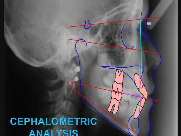
Dolphin Ceph Tracing Software
Three Lateral cephalometric radiographs were obtained for all edentulous patients (Group B) at the VDO following three different techniques firstly using Niswonger’s method (Group B1), followed by Phonetics method (Group B2), and the Swallowing threshold method (Group B3), subsequently. Group B1: Niswongers method – Cephalogram 1“Vertical Dimension at rest (VDR) =Vertical Dimension at occlusion (VDO) + Free-way space” was used to evaluate the vertical dimension at occlusion for the first cephalogram taken with Niswonger’s physiological method for all edentulous patients. Point marked adhesive triangles were placed on tip of the nose and the most prominent part of the chin. Pre-adjusted occlusion rims were then placed in the mouth.
The patients were asked to swallow and relax, a freeway space of 2mm was confirmed at this position and the VDR was recorded between the points by means of a calliper. Lateral cephalogram was made later with the rims in contact at the VDO. Group B3: Swallowing Threshold method – Cephalogram 3The upper and lower occlusion rims were placed in the mouth with cones of Aluwax placed on top of the lower occlusion rim, and the patient was requested to swallow several times. As the patient swallowed, the soft wax was reduced to the vertical dimension at occlusion.
Cephalogram was taken at Vertical dimension of occlusion by placing upper and lower record base and patient was instructed to slightly touch their rims during exposure. The radiographic unit used for digital exposures in all the study groups was Digital Cephalogram apparatus ROTOGRAPH EVO, with the recommended settings of 72 kV and 6 mAS in 4.5sec for single exposure. All digital images were then compressed in JPEG image (jpg) format and imported into the tracing software. All images were of good quality and had no artifacts that might interfere with the location of the anatomical points. Cephalometric analyses were performed with the aid of a cephalometric software program indigenously developed by our research and development team at Kamineni Institute of Dental Sciences (PRO-CEPH V3). For the computerized measurements, direct digital images were imported to the PRO-CEPH V3 software program which automatically generates measurements after digitizing a set of landmarks.

Angles Measured in Hard Tissue Inferior Gonial Angle (IGA) -The Inferior Gonial angle measures the lower facial height from the Gonion point to the Anterior Nasal Spine and Anterior Nasal Spine to the Menton.Gonial Angle (GA) – Gonial angle measured from the Articulare to the Gonion point and Gonion point to the Menton.The cephalograms were then digitised in JPEG format and loaded into the PRO-CEPH V3 software. The various landmarks were identified and marked. Linear and the required angular measurements were then tabulated by the software and recorded.
Ceph Tracing Points
ResultsIn the present study we compared the measurements of lower facial height (LFH) determined by Niswonger’s, phonetics and swallowing threshold method by linear measurement after identifying the landmarks through the PRO-CEPH V3 Software. The lower facial height, given by the height between Anterior Nasal Spine and Menton (ANS-M) recorded the value of Vertical Dimension of Occlusion (VDO).
In this study, the mean value of VDO was 46.3mm (SD 4.3) with swallowing threshold method (Sub Group B3:n=25), 45.11mm (SD 3.0) with phonetics method (Sub Group B2:n=25) and 44.51mm (SD 4.5) with Niswonger’s method (Sub Group B: n=25). In this study, we calculated the proportion between the Middle Facial Height (MFH) and Lower Facial Height (LFH), measured between Nasion (N) and Anterior Nasal Spine (ANS), and Anterior Nasal Spine and Menton (Me) respectively. We found that there was stability in the skeletal VDO, corroborating that the proportion of 0.75 was present between MFH (N-ANS) and LFH (ANS-Me).
No significant difference was found in middle and lower third ratio when comparing the measurements with Niswonger’s method (Subgroup-B1), phonetics (Subgroup-B2) and swallowing threshold method (Subgroup-B3). In dentate patients the proportion between the middle and lower thirds of the face was found 0.75 and no significant difference was found. The lower facial height is also given by the angle between Anterior Nasal Spine, Gonial point and Menton.
(ANS-Go-M) described as the Inferior Gonial Angle (IGA). We compared the angular measurement values of IGA determined by Niswonger’s, phonetics and swallowing threshold method. The mean value of IGA for Niswonger’s method was 45.71 (SD 3.4) and 46.31 (SD 4.0) with Phonetics method and 47.61 (SD 3.8) with Swallowing threshold method. Swallowing method was, on average, 1.9 degree higher than IGA obtained with the Niswonger’s method and 1.3 degree higher than the Phonetics method. Statistically insignificant difference was found between Niswonger’s and Phonetics method (p0.05).
Statistically significant differences were found between Niswonger’s and Swallowing threshold methods (p. DiscussionCephelometric analysis of determing the vertical dimension in edentulous patients have been proven to be the most accurate and acceptable method till date. Developments in computer technology have led to increasing use of digital systems both for tracing and analysing cephalometric films.
Manual Cephalometric analysis is prone to errors in identification and measurements and requires more time for calculations. Computer Assisted-Cephalometric analysis however, improves the diagnostic value of cephalometry by reducing errors and saving time.
It has been proved that Computerized cephalometry is a “smart service” which has revolutionized the dental profession providing precision and sophistication in clinical work. Earlier studies revealed that computer-aided cephalometric analysis does not introduce greater measurement error than hand tracing, as long as landmarks are identified manually (Gravely and Benzies & Enlow and Hans) ,. Computerized or Computer-Aided Cephalometric analysis eliminates the mechanical errors introduced by calibrated instrumentation. Computerized cephalometric analysis uses manual identification and location of anatomical or constructed landmarks points on digitized cephlogram image and then transfers it to computer software which completes the cephalometric analysis by automatically measuring distances and angles with respect to their corresponding landmarks. In this study the software program, PRO-CEPH V3,was used to assess, evaluate and compare the lower facial height and therefore the vertical dimension at occlusion recorded by three physiological methods, Niswonger’s, Phonetics and Swallowing Threshold.
Ceph Tracing Software
It was also used to investigate the possibility of predicting or correlating the clinical VDO through a linear measurement from the MFH:LFH proportion and through an angular measurement of the Inferior Gonial Angle (IGAm) from the measured Gonial Angle (GA) and that calculated from a regression formula: IGAc=0.508 x ( GA-15.7) by computer assisted cephalometric means. C Millet et al.in their study compared the swallowing technique with Niswonger’s method and found that the VDO determined with the swallowing method was, on average, 2.0 mm higher than VDO obtained with the use of Niswonger’s method. However, there were no statistically significant differences in their study. In our study the VDO determined with the Swallowing method (Sub Group B3) was on average 1.2 mm higher than VDO obtained with the use of phonetics method (Sub Group B2) and 1.8 mm more from Niswonger’s method (Sub Group B1).
Significant difference was found between Niswonger’s and Swallowing threshold method. ConclusionCephalometric study of the subjects with dentulous group included in this study provided important and specific parameters for optimal reconstruction of vertical dimension of occlusion in the treatment of edentulous patients. The indigenously developed software PRO-CEPH V3 is capable of making both the linear and angular measurement and therefore provide with relative credibility information regarding the possible VDO in the edentulous patients through cephalometric radiography. The use of the digital software minimizes the manual errors and provides precicional values of measurements in determining vertical dimensions in edentulous patients.
Choosing a smart way to work saves clinician time as well as provides sophistication at clinical work.


